Lymph node metastasis -- the spread of cancer from its primary site to the lymph nodes of the neck -- is one of the most critical factors determining prognosis in head and neck cancer. The presence, number, and extent of involved nodes can shift a patient's survival odds dramatically and dictate the intensity of treatment required. Neck dissection surgery, the systematic removal of lymph nodes from defined anatomical compartments, remains the cornerstone of managing cervical nodal disease. At THANC Hospital in Chennai, Dr. Vidhyadharan Sivakumar performs the full range of neck dissection procedures -- from targeted selective dissections that preserve critical nerves and structures to comprehensive radical dissections for advanced disease -- drawing on more than 3,000 head and neck surgeries and international training across 8 countries.
Understanding Lymph Node Metastasis in Head and Neck Cancer
The neck contains approximately 300 lymph nodes organised into well-defined levels (I through VII). These nodes form a drainage network that filters lymphatic fluid from the oral cavity, pharynx, larynx, thyroid, salivary glands, and skin of the head and neck. When a primary cancer develops in any of these sites, malignant cells can enter the lymphatic system and establish secondary deposits in regional lymph nodes -- this process is cervical lymph node metastasis.
India faces an outsized burden of head and neck cancer, accounting for nearly one-third of all male cancers in the country. GLOBOCAN estimates place India among the nations with the highest incidence worldwide, with over 200,000 new head and neck cancer cases annually. The prevalence of tobacco chewing, bidi smoking, areca nut use, and alcohol consumption -- particularly in southern states including Tamil Nadu -- fuels this epidemic. A significant proportion of these patients present with palpable neck nodes at diagnosis, reflecting both the biological aggressiveness of tobacco-related cancers and delays in seeking medical attention.
The status of cervical lymph nodes is the single most important prognostic factor in head and neck squamous cell carcinoma. A patient with a single ipsilateral involved node sees their survival approximately halved compared to a node-negative patient with the same primary tumour. The presence of extranodal extension -- cancer breaching the node capsule into surrounding soft tissue -- further worsens prognosis and mandates adjuvant chemoradiation. Understanding this biology underscores why meticulous neck dissection by an experienced head and neck surgical oncologist is not optional but essential. For a deeper understanding of staging, read our guide on head and neck cancer stages explained.
Types and Classification of Neck Dissection
Neck dissection has evolved substantially from the original radical procedure described by George Crile in 1906. Modern classification, standardised by the American Head and Neck Society, recognises three principal categories:
Radical Neck Dissection (RND) removes all lymph node groups from levels I through V along with three non-lymphatic structures: the sternocleidomastoid muscle, the internal jugular vein, and the spinal accessory nerve. Once the standard for all nodal disease, it is now reserved for bulky, fixed nodal disease encasing these structures.
Modified Radical Neck Dissection (MRND) removes levels I through V but preserves one or more of the three non-lymphatic structures. It is the most common therapeutic dissection when multiple node levels are involved but structures are not directly invaded.
Selective Neck Dissection (SND) removes only specific lymph node levels based on the known drainage patterns of the primary tumour. Common variants include supraomohyoid dissection (levels I-III) for oral cancers and lateral neck dissection (levels II-IV) for laryngeal and pharyngeal cancers.
| Feature | Selective Neck Dissection | Modified Radical Neck Dissection | Radical Neck Dissection |
|---|---|---|---|
| Lymph node levels removed | Selected levels (e.g. I-III or II-IV) | All levels (I-V) | All levels (I-V) |
| Spinal accessory nerve | Preserved | Usually preserved | Sacrificed |
| Internal jugular vein | Preserved | Usually preserved | Sacrificed |
| Sternocleidomastoid muscle | Preserved | Usually preserved | Sacrificed |
| Primary indication | Elective or limited nodal disease | Therapeutic for confirmed metastasis | Bulky/fixed nodal disease encasing structures |
| Shoulder morbidity | Minimal | Low to moderate | Significant |
| Typical operating time | 1-2 hours | 2-3 hours | 2-4 hours |
| Hospital stay | 3-5 days | 4-6 days | 5-7 days |
Sentinel node biopsy represents the most targeted approach, identifying and removing only the first-echelon drainage node(s) using radiotracer and/or blue dye mapping. When the sentinel node is negative on pathological examination, formal neck dissection may be avoided entirely. Dr. Vidhyadharan offers this technique for selected early-stage oral cavity cancers and head and neck skin cancers where the clinical neck is negative.
Causes and Risk Factors for Cervical Lymph Node Metastasis
The likelihood of lymph node metastasis depends on features of the primary tumour and patient-related factors:
- Primary tumour size and depth of invasion -- larger, deeper tumours have exponentially higher rates of nodal spread. For oral tongue cancer, depth of invasion greater than 4 mm significantly increases the risk and guides the decision for elective neck dissection.
- Primary site -- supraglottic laryngeal cancer and base of tongue cancer have nodal metastasis rates of 40-60% at presentation. Floor of mouth and buccal cancers also carry high risk. Glottic cancer has low nodal rates due to sparse lymphatic drainage.
- Histological grade -- poorly differentiated tumours spread more aggressively than well-differentiated ones.
- Perineural invasion and lymphovascular invasion on biopsy signal higher metastatic potential.
- Tobacco and alcohol use -- India-specific risk factors that contribute to both primary cancer development and the aggressiveness of disease.
- Delayed presentation -- a major factor in India, where patients often present with advanced-stage disease and established nodal metastasis.
- Immunosuppression -- transplant recipients and HIV-positive individuals face increased risk of aggressive head and neck cancers with early nodal spread.
Signs and Symptoms
Cervical lymph node metastasis often announces itself through palpable changes in the neck, though some patients harbour occult (clinically undetectable) disease:
- A new, persistent neck lump -- typically firm, non-tender, and progressively enlarging over weeks. This is the most common presenting sign.
- Multiple matted nodes -- several enlarged nodes that feel fused together, suggesting extranodal extension.
- Fixed mass -- a node that does not move freely, indicating fixation to underlying structures.
- Skin changes -- redness, warmth, or ulceration overlying a node in advanced cases.
- Symptoms of the primary tumour -- hoarseness, difficulty swallowing, non-healing mouth ulcer, ear pain, or nasal obstruction may coexist.
- Systemic symptoms -- unexplained weight loss, fatigue, and night sweats in advanced disease.
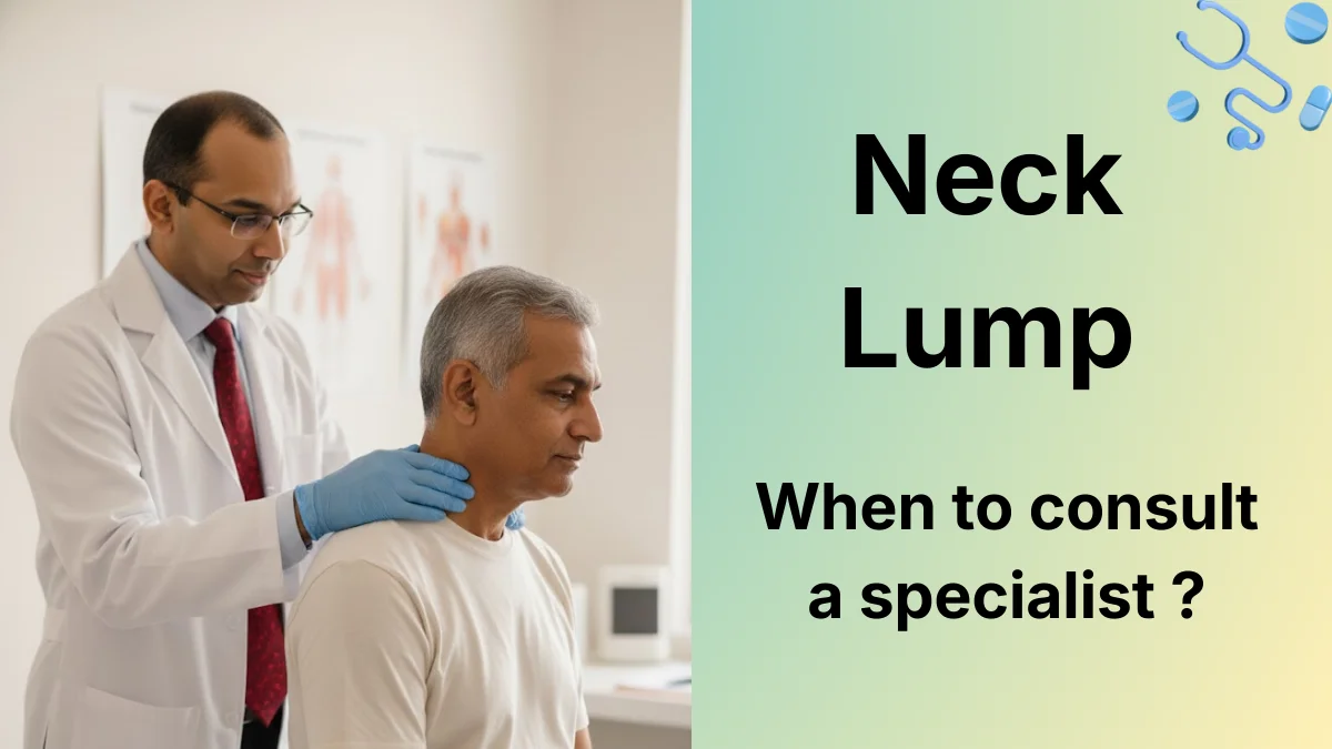
A key warning: any neck lump persisting beyond three weeks in an adult, especially a tobacco or alcohol user over 40 years, demands urgent specialist evaluation. Our detailed guide on when to worry about a neck lump helps patients understand the red flags.
Diagnosis at THANC Hospital
At THANC Hospital, Dr. Vidhyadharan employs a systematic diagnostic approach to accurately characterise cervical lymph node disease and identify or confirm the primary tumour:
- Clinical examination -- methodical palpation of all neck levels, assessment of node size, consistency, and mobility, combined with examination of the oral cavity, oropharynx, larynx, and nasopharynx.
- Ultrasound-guided FNAC -- the initial investigation for a suspicious neck node. Ultrasound identifies abnormal nodal morphology, and FNAC provides cytological diagnosis. Core needle biopsy is used when FNAC is inconclusive.
- Contrast-enhanced CT scan of the neck and chest -- defines node number, size, location, extranodal extension, and screens for pulmonary metastasis.
- MRI of the neck -- superior soft tissue resolution for evaluating extranodal extension and carotid encasement.
- PET-CT scan -- essential for staging advanced disease and detecting occult primary tumours.
- Panendoscopy with directed biopsies -- examination under anaesthesia with biopsies of any suspicious mucosal abnormality.
- p16 and HPV testing -- to identify HPV-driven cancers with distinct prognostic implications.
Every case is discussed at the multidisciplinary tumour board comprising the head and neck surgeon, radiation oncologist, medical oncologist, radiologist, pathologist, and rehabilitation specialists to formulate a consensus treatment plan.
How Dr. Vidhyadharan Treats Lymph Node Metastasis
Cervical lymph node management is always integrated with primary tumour treatment. Dr. Vidhyadharan's MCh from Amrita Institute, European Board certification (FEB-ORL HNS), and training across 8 countries equip him to execute the optimal neck dissection strategy for every scenario.
Elective Neck Dissection (Clinically Node-Negative Neck)
When imaging and clinical examination show no evidence of nodal disease but the primary tumour carries a significant occult metastasis risk (generally exceeding 15-20%), Dr. Vidhyadharan performs elective selective neck dissection. For oral cavity cancers, this typically involves a supraomohyoid dissection (levels I-III). For laryngeal and pharyngeal primaries, a lateral dissection (levels II-IV) is standard. Sentinel node biopsy is offered as an alternative for early oral cancers, potentially sparing patients a formal dissection when the sentinel node is negative.
Therapeutic Neck Dissection (Clinically Node-Positive Neck)
When imaging or FNAC confirms nodal metastasis, therapeutic neck dissection is performed -- modified radical dissection preserving key structures wherever oncologically safe, or radical dissection when structures are encased by tumour. Dr. Vidhyadharan's training at Amrita Institute established his foundation in nerve-sparing technique, further refined during international fellowships.
Salvage Neck Dissection After Chemoradiation
Patients who undergo primary chemoradiation for advanced head and neck cancer and develop persistent or recurrent nodal disease require salvage neck dissection. This is among the most technically demanding operations due to radiation-induced fibrosis and altered tissue planes. Dr. Vidhyadharan's extensive experience with salvage surgery -- honed through managing recurrent cancers at THANC Hospital -- ensures safe dissection through scarred, irradiated fields.
Combined Procedures
Neck dissection is commonly performed simultaneously with primary tumour resection (composite resection) and immediate reconstruction using local, regional, or free flaps. Dr. Vidhyadharan's microsurgical training at Chang Gung Memorial Hospital, Taiwan, enables him to perform complex reconstructions following extensive resections that include neck dissection, minimising the number of anaesthetic episodes and overall treatment duration.
Adjuvant Therapy After Neck Dissection
Pathological findings from the neck dissection specimen guide adjuvant therapy decisions. The presence of extranodal extension, positive margins, multiple positive nodes, or perineural invasion may mandate adjuvant radiation or concurrent chemoradiation. These decisions are made through the THANC Hospital tumour board.
What to Expect: Your Treatment Journey
Dr. Vidhyadharan and the THANC Hospital team ensure patients understand every step of their neck dissection journey:
Week 1 -- Evaluation and staging: Comprehensive clinical examination, imaging (CT, MRI, PET-CT as indicated), ultrasound-guided FNAC or core biopsy, and panendoscopy if the primary is not yet identified. Most investigations are completed within the first week.
Week 2 -- Tumour board and treatment planning: Your case is presented to the multidisciplinary tumour board. Dr. Vidhyadharan explains the recommended dissection type, expected functional outcomes, need for combined primary tumour surgery and/or reconstruction, and adjuvant therapy plan. This consultation is unhurried, and patients are encouraged to bring family members and prepare questions.
Week 2-3 -- Pre-operative preparation: Pre-anaesthesia evaluation, blood work, cardiac and pulmonary clearance if needed, nutritional optimisation, and pre-operative physiotherapy assessment to establish baseline shoulder function.
Surgery day: Neck dissection typically takes 1.5 to 4 hours depending on type and whether it is combined with primary tumour surgery and reconstruction. General anaesthesia is used throughout. A suction drain is placed in the neck at the end of the procedure.
Hospital stay (3-7 days): Drain monitoring, wound care, pain management, early mobilisation, and shoulder physiotherapy. Drains are removed when output falls below 30 ml per day (typically 3-5 days).
Post-discharge rehabilitation: Shoulder physiotherapy continues for 6-12 weeks. Follow-up at 2 weeks, 6 weeks, 3 months, then every 3 months for two years, every 6 months for years 3-5, and annually thereafter.
Recovery and Rehabilitation
Recovery after neck dissection depends on the type and extent of surgery performed:
Selective neck dissection patients experience the fastest recovery. The spinal accessory nerve is preserved, shoulder function remains intact or mildly affected, and most patients return to normal activities within 2-3 weeks. Neck stiffness and mild numbness of the ear and neck skin are common but temporary.
Modified radical neck dissection patients may experience temporary shoulder weakness and reduced range of motion, even when the spinal accessory nerve is preserved, due to surgical traction. Dedicated physiotherapy targeting shoulder abduction and overhead reach is essential. Most patients recover near-normal shoulder function within 3-6 months.
Radical neck dissection patients face the greatest rehabilitation challenge. Sacrifice of the spinal accessory nerve results in permanent shoulder droop and limited abduction. Intensive physiotherapy helps compensate by strengthening the trapezius alternatives (levator scapulae, rhomboids) and maintaining range of motion. Loss of the sternocleidomastoid muscle alters neck contour cosmetically.
Common post-operative experiences include numbness of the neck skin and ear (expected, partially recovers over months), mild neck tightness, and transient facial or submental lymphoedema. Chyle leak from thoracic duct injury during left-sided dissection is managed conservatively with dietary modification. THANC Hospital's rehabilitation programme integrates physiotherapy, speech-language pathology, and nutritional support.
Outcomes and Prognosis
The impact of neck dissection on survival is well documented. Adequate surgical clearance of nodal disease, combined with appropriate adjuvant therapy, significantly improves outcomes:
- Node-negative patients undergoing elective neck dissection have excellent regional control rates exceeding 95%, with the dissection primarily serving a staging role and preventing regional recurrence.
- Single node, no extranodal extension -- adjuvant radiation achieves regional control in 90-95% of cases.
- Multiple positive nodes or extranodal extension -- adjuvant chemoradiation improves regional control to 80-85%, though overall prognosis is significantly impacted.
- Salvage neck dissection after chemoradiation failure achieves regional control in 50-70% of carefully selected patients.
The number of positive nodes, presence of extranodal extension, and the level of involved nodes all influence prognosis. High-volume surgeons who perform meticulous, complete dissections with adequate node yields consistently demonstrate better regional control rates in the literature. Dr. Vidhyadharan's 3,000+ head and neck surgeries reflect this volume-outcome relationship.
Why Choose Dr. Vidhyadharan at THANC Hospital
Neck dissection demands precision in a region dense with critical nerves and blood vessels. Dr. Vidhyadharan Sivakumar brings:
- MCh (Head & Neck Surgery), Amrita Institute of Medical Sciences -- India's premier super-specialty programme for head and neck surgical oncology.
- European Board certification (FEB-ORL HNS) -- among the few Indian surgeons holding this credential.
- Training across 8 countries -- including Canada (Toronto General Hospital), Australia (Royal Adelaide Hospital), and Taiwan (Chang Gung Memorial Hospital), each adding layers of surgical expertise.
- 3,000+ head and neck surgeries -- providing the volume that published evidence associates with better outcomes.
- Co-editor, "Comprehensive Management of Head and Neck Cancer" (Jaypee Brothers, 2021) -- contributing to the educational foundation of the specialty.
- Nerve-sparing philosophy -- preservation of the spinal accessory nerve, marginal mandibular nerve, and hypoglossal nerve wherever oncologically safe, using intraoperative monitoring and meticulous technique.
THANC Hospital provides the infrastructure to match this expertise: advanced imaging, intraoperative nerve monitoring, a dedicated head and neck cancer centre, and a multidisciplinary team focused exclusively on head and neck oncology.
To schedule a consultation with Dr. Vidhyadharan Sivakumar, call +91 73059 53378 or request an appointment online.